
What Happens If a CT Scan Shows an Abnormal Spot in the Lung?
What Happens If a CT Scan Shows an Abnormal Spot in the Lung?


What Happens If a CT Scan Shows an Abnormal Spot in the Lung?
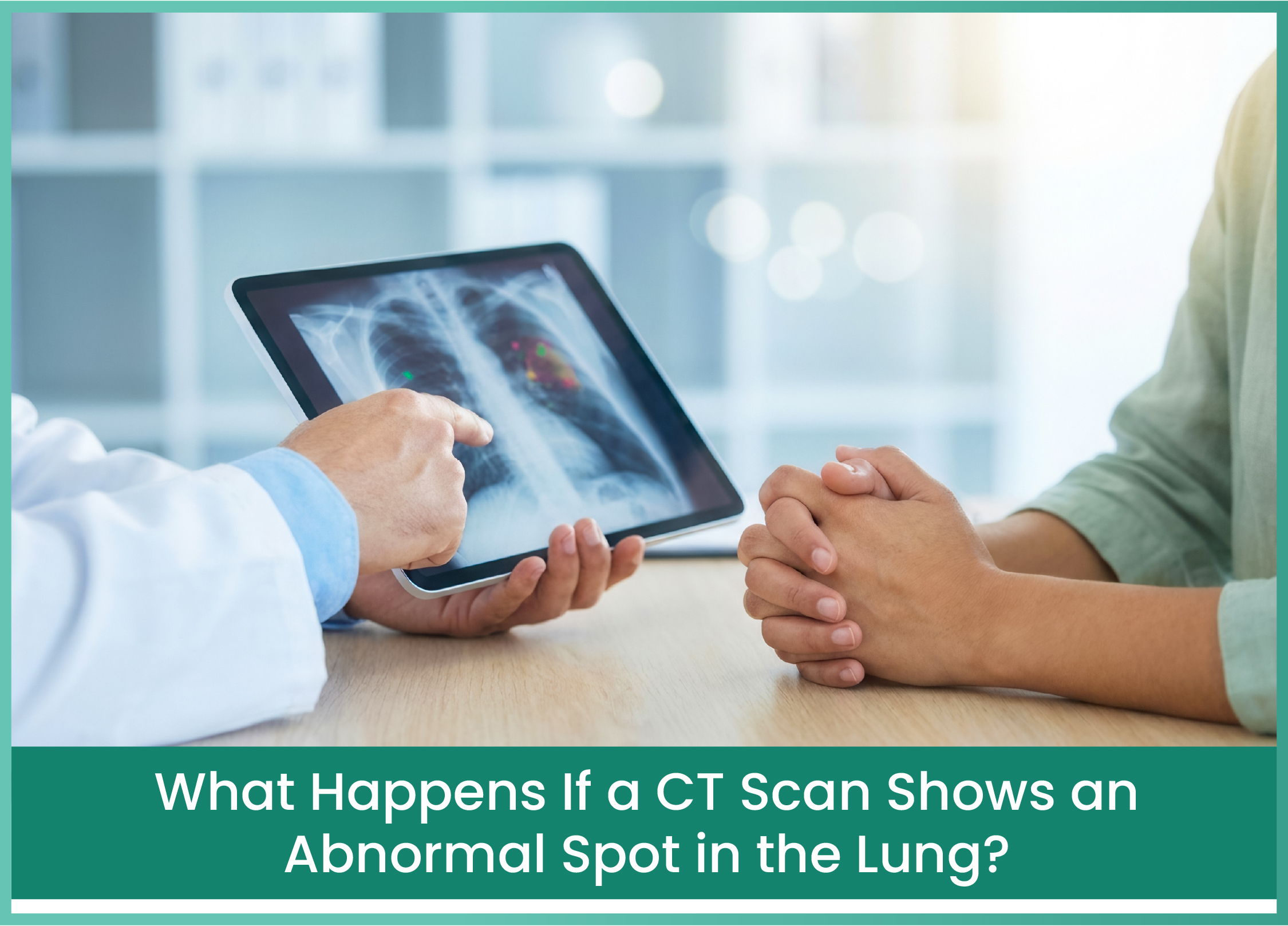
Getting a CT scan report that mentions an abnormal spot in the lung can be an unsettling experience. For many people, the first thought that comes to mind is lung cancer. Questions such as "Is it dangerous?", "Could it be an infection?" or "Will I need a biopsy?" can create anxiety, especially when the report contains unfamiliar medical terms.
However, an abnormal spot in the lung does not always mean cancer. In many cases, it may be related to an old infection, inflammation, tuberculosis or a small area of scar tissue left behind after a previous lung condition. Sometimes, these spots are discovered accidentally when a person undergoes a CT scan for an entirely different medical reason.
A CT scan provides a detailed picture of the lungs and helps doctors identify changes that may not be visible on a regular chest X-ray. These changes can appear as small nodules, hazy patches, areas of increased density or larger abnormalities within the lung tissue. Although some findings are harmless, others may need closer examination to understand their cause.
The important thing is to avoid jumping to conclusions after reading the scan report. Doctors consider several factors, including the size, shape, location and appearance of the abnormality, before deciding whether further investigations are necessary. Understanding what an abnormal lung spot means and what happens next can help patients approach the situation with greater confidence.
Why Does a CT Scan Show an Abnormal Spot in the Lung?
The lungs contain millions of tiny air sacs that help the body absorb oxygen and remove carbon dioxide. Healthy lung tissue has a characteristic appearance on a CT scan. When an area becomes inflamed, develops scar tissue or contains an unusual growth, it may appear different from the surrounding lung tissue.
Radiologists use terms such as pulmonary nodule, lung opacity, ground-glass opacity and lung lesion to describe these findings. Although these terms may sound frightening, they simply describe the appearance of an abnormal area on the scan. They do not independently confirm a disease.
For example, a pulmonary nodule is a small, rounded area of tissue within the lung. It may develop following an infection or due to a benign growth. In some cases, however, it may represent an early lung cancer. Similarly, a ground-glass opacity describes a hazy area in which the underlying lung structures can still be partially seen. This appearance can occur due to infection, inflammation and certain lung cancers.
The reason behind an abnormal finding can only be understood after considering the patient's medical history, symptoms and imaging characteristics. This is why a doctor may recommend reviewing previous scans or undergoing additional investigations rather than immediately starting treatment.
Can an Abnormal Spot in the Lung Be Caused by an Old Infection?
One of the common reasons for an abnormal lung finding is a previous respiratory infection. Conditions such as pneumonia and tuberculosis can cause inflammation and damage to lung tissue. Even after the infection has been successfully treated, some changes may remain visible on imaging.
For instance, a person who had tuberculosis several years ago may have small scars or calcified nodules in the lungs. These findings may remain even when there is no active infection. Similarly, pneumonia can sometimes leave temporary areas of inflammation that gradually improve over time.
This is particularly relevant in India, where tuberculosis continues to be an important cause of lung disease. A person with a history of TB may understandably become concerned when a new CT scan shows an abnormal spot. However, the finding does not automatically mean that tuberculosis has returned or that cancer has developed.
Doctors usually compare the current scan with older imaging reports to understand whether the abnormality is new, unchanged or increasing in size. If the appearance suggests an infection, additional tests may be recommended to identify the underlying cause.
When Can an Abnormal Lung Spot Be a Sign of Lung Cancer?
Although many lung abnormalities are non-cancerous, some may require investigation for lung cancer. Lung cancer develops when abnormal cells in the lung begin multiplying uncontrollably and form a growth.
In certain patients, the disease may initially appear as a small pulmonary nodule. These nodules may be discovered during routine imaging even when the person has no cough, chest pain or breathing difficulty.
Doctors pay attention to certain characteristics that may increase suspicion. These include an irregular shape, spiculated borders, increasing size or changes in the density of the nodule. A history of smoking, older age and previous cancer may also influence the assessment.
However, none of these factors alone can establish a cancer diagnosis. Even a suspicious-looking nodule may have a non-cancerous explanation. When further evaluation suggests the possibility of lung cancer, investigations such as PET-CT imaging or a biopsy may be recommended.
A timely consultation with an appropriate specialist can help patients understand the findings and avoid unnecessary delays in diagnosis.
What Happens After an Abnormal Spot Is Found on a CT Scan?
The next step is not the same for every patient. Some people may only need another CT scan after a recommended interval, while others may require more detailed investigations.
The doctor first reviews the scan report and, whenever possible, examines the actual images. The size and location of the abnormality are considered along with other findings in the chest. Previous CT scans and chest X-rays are particularly useful because they help establish whether the spot has been present for a long time or has developed recently.
The patient's medical history also plays an important role. A person who recently recovered from pneumonia may have a different explanation for an abnormal lung opacity than someone with a growing nodule and a significant smoking history.
Depending on the findings, the doctor may recommend monitoring, additional imaging or a tissue biopsy. The purpose of these investigations is to identify the cause of the abnormality and determine whether treatment is necessary.
Patients who have concerning findings or a confirmed diagnosis of lung cancer may benefit from consulting a lung cancer specialist in Mumbai. A medical oncologist can help assess the diagnosis, understand the available treatment options and coordinate further cancer care.
Does Every Abnormal Lung Nodule Need a Biopsy?
No. A biopsy is not necessary for every abnormal spot detected on a CT scan.
Small nodules with reassuring characteristics may only require periodic monitoring. In these situations, doctors may recommend a follow-up CT scan to check whether the nodule changes over time.
However, when an abnormality becomes larger, develops suspicious features or shows other concerning changes, further investigations may be required. A biopsy helps doctors collect a small tissue sample and examine it under a microscope to determine whether cancer cells are present.
The decision depends on the patient's individual circumstances. Doctors consider the potential benefits of obtaining a tissue diagnosis against the risks of the procedure.
This is why patients should not assume that a biopsy is always required after an abnormal CT scan. The most appropriate investigation is the one that answers the medical question safely and accurately.
Why Timely Medical Evaluation Matters?
An abnormal spot in the lung can have several explanations, and the majority of small pulmonary nodules are not cancerous. However, ignoring the finding is not advisable.
Some lung cancers can develop without producing noticeable symptoms during their early stages. A person may feel completely healthy while an imaging scan reveals an abnormality that requires further assessment.
Following the recommended investigation schedule allows doctors to identify changes and decide whether additional evaluation is needed. If lung cancer is confirmed, determining its type and stage helps guide treatment planning.
For patients seeking guidance from Dr. Reetu Jain, a medical oncologist in South Mumbai, understanding the CT scan findings, previous medical history and recommended investigations can help establish the appropriate next steps in cancer evaluation and care.